SILVER STAINING
Our excellent silver stain is based on the ammoniacal silver/formaldehyde method of Oakley et. al. (Anal. Biochem.105: 361, 1980). This method involves preliminary glutaraldehyde treatment of the slab gel to fix proteins by cross linking. The pre-treatment also adds glutaraldehyde side chains to the proteins, increasing sensitivity since these groups are sites for silver deposition (Dion A, and Pomenti A, Anal. Biochem. 129:390, 1983). We find this method to be >10 times more sensitive than Coomassie blue staining, depending on the protein, even though less protein is loaded. Generally, 50 ng of purified protein gives a highly visible spot; see our internal standard (tropomyosin) marked with a black arrow on example gels. However, some proteins that are detectable with Coomassie don’t stain at all with silver. Although very sensitive, this method gives semi-quantitative rather than quantitative results. The linear range for plots of stain density versus ng protein varies from protein to protein with different saturation levels. Day to day variability in stain and background intensity occurs. Even so, silver staining is useful for computerized comparisons because spot values are normalized – expressed as a percentage of all spots combined. Values from duplicate gels are always averaged. Silver staining often interferes with mass spectrometry but the 2D patterns match well to a heavily loaded Coomassie-stained duplicate gel. Any spot present on the latter, no matter how faint, is within range for mass spectrometry for protein identification.
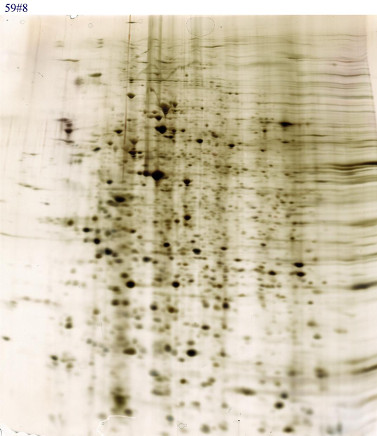
We offer both standard silver staining and special silver staining for mass spectrometry. Follow the link for pricing details.
Call 800-462-3417 or email 2d@kendricklabs.com for a price quote without obligation.