• Skip to primary navigation
  • Skip to main content
  • Skip to primary sidebar
  • Skip to footer

Three Decades of Experience in
Protein Contract Research
Request a Quote
  • Home
  • HCP Analysis
    • HCP Overview
    • Host Cell Protein (HCP) Antibody Analysis Method
    • Host Cell Protein (HCP) Results & Report
    • HCP Pricing
    • Product Characterization
    • Drug Substance Testing
  • 2D
    • 2D Overview
    • 2D Catalog
    • 2D Computer Comparisons
    • 2D Examples
    • Coomassie Blue Staining
    • Silver Staining
    • Standardization
    • Sample ID Form (PDF)
  • WB
    • 1D & 2D Western Blotting Services
    • HCP Analysis WB
    • Acetyl-lysine WB
    • P-Ser/P-Thr WB
    • P-Tyrosine WB
  • 1D
    • 1D Catalog
    • Food/Nutrition Testing
    • Sample ID Form (PDF)
  • Support
    • Sample Preparation
    • Accreditation
    • FAQs
    • Kendrick Labs Citations
    • White Papers/Recent Publications
    • Presentations
    • Misc
    • Terms & Conditions
  • Contact
    • Partnership Opportunities
    • Contact Us
    • How to Send Samples
    • Mission and People
    • Careers at Kendrick Labs
    • Sitemap

HCP Testing of Drug Substance and In-Process Samples

2D SDS PAGE analysis of purified Drug Substance (DS) samples provides an orthogonal method of confirming HCP removal during processing steps. The analysis steps are similar to those used for testing HCP antibodies on whole cell lysates. Samples are loaded on duplicate 2D silver-stained gels and on duplicate 2D gels to be transferred to PVDF for western blotting. One of the western blots is probed with the anti-HCP antibody. The duplicate western blot is probed with an antibody against the DS, since the sample contains a large amount of DS this helps determine which proteins are DS and which are HCP. When determining the HCP antibody coverage, the DS spots are omitted from the analysis.
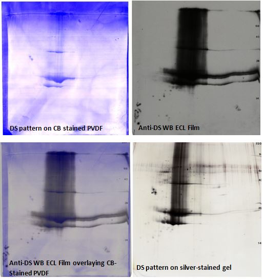
Anti-drug substance 2D western blot: stained PVDF, anti-DS 2D film, overlay image, 2D gel Figure 1. Anti-DS 2D western blot images. Upper Left: Coomassie blue-stained PVDF, Upper Right: anti-DS ECL Western blot film, Lower Left: Western blot overlay image; and Lower Right: silver-stained 2D gel pattern. Spots reacting to the DS ab are omitted from the analysis. Heavily loaded DS samples sometimes show vertical trailing, breakdown products, and polymers as seen here.

Film images are warped to the silver-stained 2D gel with TotalLab SameSpots software using the Coomassie blue-stained PVDF membrane image (before western blotting) to facilitate alignment. Spots detected by the anti-DS ab are omitted from the analysis along with any buffer blank spots. The remaining spots detected by silver-staining and the anti-HCP ab are outlined to determine the anti-HCP ab coverage. Spots visible only with the anti-HCP ab but not detectable by silver staining are added to the analysis as indicated by small blue dots on the silver-stained gel.
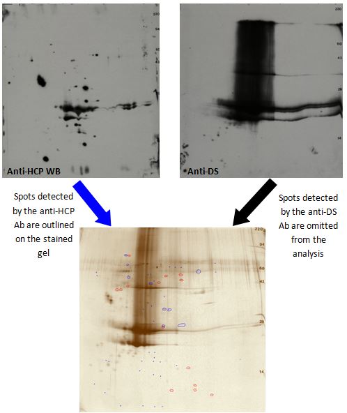
2D Gel analysis image excludes buffer blank & drug substance spots. HCP ab detects 75% of HCP spots Figure 2. 2D Gel Difference Image showing silver-stained gel spots detected by the anti-HCP ab in blue, spots not detected by the antibody in red, and spots detected by the anti-DS ab not outlined (buffer blank spots also omitted). The anti-HCP ab detected 41 spots out of the total 55 HCP spots in the analysis (75%).

This method can be used to monitor the removal of HCPs during the DS purification process. It is also useful to confirm removal of most HCPs from the final DS. Finally, in combination with duplicate Coomassie blue-stained 2D gels, HCPs can be further analyzed using mass spectrometry.
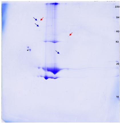
Mass spectrometry compatible Coomassie blue-stained 2D gel. Candidates ID by LC/MS marked by arrowsFigure 3. Coomassie blue-stained 2D gel compatible with mass spectrometry. Spot candidates for identification by mass spectrometry are marked with blue (detected by HCP antibody) or red (not detected by HCP antibody) arrows.

Identification of protein spots of interest by mass spectrometry would give additional supporting information. Spots not detected by the HCP antibody or DS antibody could be DS breakdown products, DS multimers, or HCP not detected by the ab. Determining the identity of HCP proteins (either detected or not by the HCP ab) allows for assessment of importance of their removal from the final DS and informs strategies for modification of the purification process for removal of remaining HCPs.

Primary Sidebar

Contact Us

  800-462-3417
 2d@kendricklabs.com
 1202 Ann St, Madison, WI 53713

Key Links

  • 2D Services and Pricing
  • 1D Services and Pricing
  • Download Catalog (pdf)
  • 2D SDS Compatibility
  • Submit Samples
  • Sample Preparation

Klabs Email List

Loading

Footer


Copyright © 2025